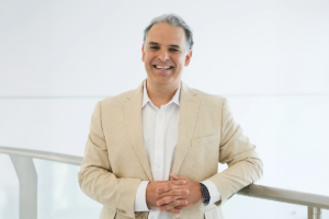
David Brenner, MD
President and CEO
Donald Bren Chief Executive Chair
Professor, Center for Metabolic and Liver Diseases
Metabolic dysfunction-associated steatohepatitis or MASH is a severe form of fatty liver disease, a condition that afflicts roughly one-third of adults worldwide: nearly 2 billion people. Untreated, MASH can lead to
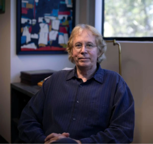
Lukas Chavez, PhD
Associate Professor
NCI-Designated Cancer Center
Childhood brain cancer is not one disease, but many, each with its own genetic fingerprint and its own way of evading treatment. Advances in genomics now allow us to read these fingerprints in extraordinary detail, uncovering the hidden instructions that make
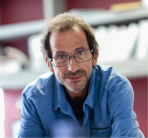
Jerold Chun, MD, PhD
Professor
Center for Neurologic Diseases
The human brain remains one of life’s greatest mysteries and challenges, being responsible for all human activities that require its myriad functions. However, our understanding of the brain remains incomplete and this deficiency is perhaps most evident through hundreds of brain
Gregg Duester, PhD
Professor
Center for Cardiovascular and Muscular Diseases
Science is facing a reproducibility crisis. In the biomedical sciences, the inability to validate and reproduce findings is slowing progress in understanding basic principles. Among the fields hindered by irreproducibility is retinoic acid research. Fortunately, new techniques provide a
Hudson Freeze, PhD
Professor and Director
William W. Ruch Distinguished Endowed Chair
My fascination with sugars began early, sparked not by science but Oreos.
As a child, I couldn’t get enough. Later came chocolate, and with it a growing sense that sugars held more than just
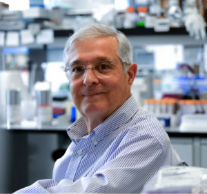
Michael Karin, PhD
Director and Professor
Center for Metabolic and Liver Diseases
Despite much progress in the past 100 years in understanding the pathogenesis of many common diseases and their treatments, we do not understand how these diseases are initiated.
For example, take pancreatic cancer. We know that
Kelly Kersten, PhD
Assistant Professor
NCI-Designated Cancer Center
I have always been fascinated by the paradoxical role of the immune system in cancer. On the one hand our immune system protects us from infections and disease. But in some cases, it can turn against us. With the recent advancements
Caroline Kumsta, PhD
Assistant Professor
Center for Cardiovascular and Muscular Diseases
What doesn’t kill you makes you stronger. It’s true not just for people, but for our cells. My research explores hormesis, the phenomenon where mild, manageable stressors such as exercise, heat or dietary changes trigger powerful defense systems
Ahmed Mahmoud, PhD
Associate Professor
Center for Cardiovascular and Muscular Diseases
Heart failure remains the leading cause of death worldwide because the human heart cannot regenerate after injury. Unlike newborn mammals, whose hearts can regrow following damage, the adult heart heals by forming scar tissue, leading to permanent loss
Jamey Marth, PhD
Professor
Center for Metabolic and Liver Diseases
I seek to discover and therapeutically control the metabolic origins of common diseases.
From human twin studies and human genome sequence comparisons, it has become evident that genetics plays a limited and minor role in the
José Luis Millán, PhD
Professor
Center for Cardiovascular and Muscular Diseases
Why do our skeleton and teeth, but not our soft organs, calcify under physiological conditions? And what goes wrong in hypophosphatasia (HPP), a condition in which children display soft bones and premature loss of teeth (and sometimes die) or
Andrei Osterman, PhD
Professor
Center for Metabolic and Liver Diseases
We live in the microbial world! Indeed, our body provides an ecological niche for myriad diverse bacteria. Among them are benign (and even beneficial) commensals comprising most notably the human gut microbiome, but also sporadic invaders, including deadly bacterial

Xueqin (Sherine) Sun, PhD
Assistant Professor
NCI-Designated Cancer Center
Our research aims to understand why cancer develops, identify its “Achilles’ heel” and ultimately create more effective treatments.
Cancer arises when cells misread or misinterpret their DNA, leading to abnormal gene activity driven by both genetic changes and

Kevin Tharp, PhD
Assistant Professor
NCI-Designated Cancer Center
I want to understand how cells adapt to different environments.
This may seem like a simple question, but cells use a complex network of interconnected sensors to determine where they are and what to do. They physically interact with their surroundings
Xiao Tian, PhD
Assistant Professor
Center for Neurologic Diseases
Aging is a mystery that has driven human curiosity since the dawn of history. Modern research is revealing the underlying molecular mechanisms that shed light on this ancient question. I’m interested in understanding how aging leads to diseases that are
Alessandro Vasciaveo, PhD
Assistant Professor
NCI-Designated Cancer Center
My goal is to develop large-scale datasets and artificial intelligence as primary scientific instruments, not simply auxiliary tools.
Instead of focusing on individual genes or pathways in isolation, we want to create programs that integrate massive, multi-modal amounts of information, including
Kristiina Vuori, MD, PhD
Professor
Pauline and Stanley Foster Distinguished Chair
NCI-Designated Cancer Center
Why do recurrent cancers often exhibit resistance not only to prior treatments, but also to unrelated therapies? What is behind this “super-resistance” and how can we overcome it?
The “cancer cell autonomous” model posits that